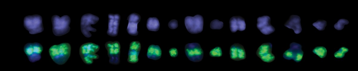
Extrachromosomal DNA (ecDNA) are circular, acentric DNA elements that carry amplified oncogenes and drive tumor progression and therapy resistance across many cancers. Because they lack centromeres, ecDNA are not subject to the machinery that partitions chromosomes evenly at mitosis. They are inherited unequally, reach copy numbers far beyond what chromosomal amplification achieves, and generate the intratumoral heterogeneity on which selection acts. Much of their oncogenic potential follows from this.
Unequal inheritance, however, is not the same as unreliable inheritance. Acentric elements should be lost from the nucleus at an appreciable rate, and ecDNA are not. How faithfully they are transmitted, and what machinery ensures it, were poorly understood.
What we found
Across a panel of ecDNA-positive cancer cell lines, sequence-specific DNA FISH in newly formed daughter-cell pairs showed that ecDNA are retained in daughter nuclei with high fidelity despite the absence of centromeres. That fidelity implied an active mechanism rather than passive partitioning.
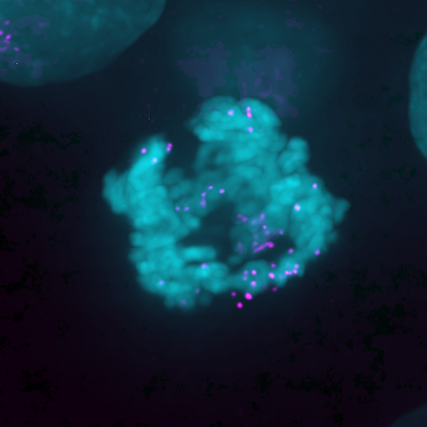
Live-cell imaging of TetO-tagged ecDNA identified it. ecDNA hitchhike on mitotic chromosomes, tracking with the chromosome ends through division. Genome-wide Hi-C confirmed the association physically, with ecDNA amplicons preferentially contacting the ends of mitotic chromosomes, and showed the contacts to be mitosis-specific rather than a carryover from interphase.
The tether depends on transcription that persists through mitosis, when most of the genome is silent. Using intron-directed RNA FISH we found that PVT1, the long non-coding RNA co-amplified with MYC on the ecDNA, continues to be transcribed during mitosis. Inhibiting mitotic transcription with triptolide disrupted ecDNA clustering, released the elements from mitotic chromosomes, and drove their mis-segregation into the cytosol, where they formed micronuclei or reintegrated into chromosomes as homogeneously staining regions.
We propose that ongoing mitotic transcription generates an RNA-based tether linking ecDNA to chromosome ends and protecting them from cytosolic loss. This accounts for how ecDNA sustain oncogene amplification across successive divisions.
Open questions
What constitutes the tether
Mitotic transcription is required and ecDNA contact chromosome ends, but the molecular link between them is unidentified. Whether the connection is made by RNA directly, by proteins recruited to nascent transcripts, or by both is unknown.
What occupies the ecDNA-proximal environment
ecDNA reside in a distinct protein neighborhood within the nucleus. Defining its composition is a prerequisite to determining which factors maintain attachment and which protect the elements from damage.
How ecDNA reintegrate to form homogeneously staining regions
Elements released from mitotic chromosomes can insert back into the genome as HSRs. This constitutes a second route to oncogene amplification and a distinct form of structural rearrangement. The mechanism is unknown.
Whether circularity is required
The circular topology of ecDNA may be functionally necessary rather than incidental. We are asking whether it is required for maintenance and for faithful segregation.
Selected publications
- Nichols A, Choi Y, Norman RX, Chen Y, Striepen J, Salataj E, Toufektchan E, Koche R, Maciejowski J. Chromosomal tethering and mitotic transcription promote ecDNA nuclear inheritance. Molecular Cell 2025;85(15):2839–2853.DOIPubMed 40614723
- Nichols A, Salataj E, Choi Y, Hamard PJ, Koche R, Maciejowski J. Protocol for the generation of low-input Hi-C sequencing libraries of FACS-isolated mitotic cells. STAR Protocols 2025;6(4):104241.DOIPubMed 41317326
- Raeisi Dehkordi S, et al., including Maciejowski J. Breakage fusion bridge cycles drive high oncogene number with moderate intratumoural heterogeneity. Nature Communications 2025;16:1497.DOIPubMed 39929823
Rotation projects in this area are listed on the Join page.